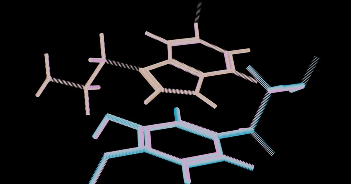
Disulfide Bonds in Peptides: How Cysteine Bridges Fold Structure
Cysteine is the only standard amino acid whose side chain can covalently link to another residue's side chain. That single bond-forming ability shapes whole families of peptides. Here is how disulfide bonds form, how they lock a peptide into a defined fold, and why the exact cysteine bridge connectivity matters so much to researchers.
by Research Assistant·
Most amino acid side chains keep to themselves. They pack, they attract, they repel — but they never form a permanent chemical bond with each other. Cysteine breaks that rule. Its sulfur-bearing side chain can join with the side chain of a second cysteine to make a genuine covalent link, and that one trick quietly shapes whole families of peptides researchers study today. Everything below is described in structural and chemical terms and is for research use only — not anything intended for human or animal use. We'll walk through what a disulfide bond is, how it locks a peptide into a defined shape, the cellular machinery that builds these bonds correctly, and why the exact pairing of cysteine bridges is one of the hardest things to get right in a disulfide-rich peptide.
What a Disulfide Bond Actually Is
In one line: a disulfide bond is a covalent bridge that joins the sulfur atoms of two cysteine residues. Everything else is detail layered on top of that idea.
From two thiols to one cystine
Every cysteine carries a thiol group — an -SH — on its side chain. Bring two of those thiols close together in an oxidizing environment, and they shed their hydrogen atoms while their sulfur atoms link directly. As the reference chemistry puts it, "disulfide bonds in proteins are formed between the thiol groups of cysteine residues by the process of oxidative folding." The joined pair even earns its own name: cystine. It helps to separate this from the bond that builds the chain itself. Running residue to residue in sequence, the peptide backbone is held together by the amide bond that forms the peptide backbone. A disulfide bond does something else entirely — it reaches across the folded chain to clamp two cysteines together, even when they sit far apart in the sequence.
Notation and scale
Since a disulfide can connect residues that are distant in the sequence, chemists label each bond by the two positions it joins, writing something like C26–C84 to name the exact pair. And this is no rare curiosity. Disulfide bonds turn up across a large share of known protein structures, and their cross-linking power shows up in ordinary material: keratin, the protein in hair and feathers, owes much of its toughness to a high disulfide content. The same chemistry that stiffens a strand of hair is what rigidifies a small research peptide.
How Disulfide Bonds Stabilize the Fold
The short version: a disulfide bond makes the correctly folded shape the easy, low-energy state — and makes coming apart harder.
Biasing the chain toward the folded topology
One bond stabilizes structure in several ways at once. Tie two segments of the chain together and you "bias the protein towards the folded topology," while also lowering the entropy of the unfolded state so the open, floppy form is less favorable from the start (Feige & Hendershot, 2011). In some peptides the bond seeds a hydrophobic core, too, pulling nearby water-avoiding residues in around it. The upshot is simple: unfolding becomes less likely, especially across otherwise labile stretches. Think of it as a spot weld holding shut a part that would otherwise flex open.
Where the environment allows it
Disulfide bonds are picky about their surroundings. They form readily in oxidizing compartments and stay unstable in reducing ones. In cell-biology terms, the oxidizing endoplasmic reticulum (ER) is where these bonds get built, while the reducing cytosol keeps them from lasting. That single fact explains why secreted and cell-surface proteins carry most of the disulfides researchers observe, and why cytosolic proteins carry so few. The ER even turns the requirement into a checkpoint: in these studies, a protein still carrying unpaired cysteines can't leave the ER, so only fully bonded, correctly matured molecules move onward. The bond isn't just structural glue — it doubles as a quality-control signal.
The Machinery Behind Oxidative Folding
Here's the key idea: in living systems, the right disulfide bonds are placed by dedicated enzymes, not left to chance collisions.
PDI and Ero1
The central catalyst is protein disulfide isomerase (PDI), a family of enzymes built around thioredoxin-like domains with a signature CXXC active-site motif that shuttles between a reduced (dithiol) and an oxidized (disulfide) form. PDI hands disulfide bonds to folding substrates, and it's re-oxidized in turn by an enzyme called Ero1, which passes electrons down to molecular oxygen and throws off hydrogen peroxide as a byproduct (Bulleid, 2012). Mammalian cells keep backups on hand — peroxiredoxin IV, glutathione peroxidases 7 and 8, and vitamin K epoxide reductase can all feed the same oxidative pathway. That redundancy tells you how much the cell depends on reliable disulfide formation.
Thiol–disulfide exchange and reshuffling
The chemistry that lets enzymes fix mistakes is thiol–disulfide exchange. A cysteine thiolate attacks an existing disulfide bond, forms a brief mixed intermediate, and swaps partners. This reshuffling runs much faster than the reactions that change the total number of bonds — which is exactly what makes efficient error-correction possible. The cell can rearrange which cysteines are paired far more quickly than it makes or removes bonds outright (disulfide biochemistry overview). Correct folding, as the ER literature stresses, takes enzymes that both introduce disulfides between nearby cysteines and take apart the ones that form during folding but don't belong in the final structure.
Native vs. Non-Native Connectivity
Same cysteines, different pairings — and you can end up with a very different molecule. That's the crux of connectivity.
Folding isomers
A cysteine-rich peptide doesn't snap straight to the right answer. Early in folding, non-native (or "scrambled") disulfide pairings coexist alongside the correct ones. Studies tracking synthetic polypeptides across a folding reaction watch the non-native species fade in the late stages as the native pattern piles up (Chang et al., 2019). The stakes here are real. In one synthetic cardiotoxin study, scrambled isomers were far less active than the correctly folded form — the correct molecule showed a clean β-sheet structure, while the mispaired versions collapsed toward random coil. Same amino acids, same number of bonds, wildly different behavior in the assay.
Finding the correct partners
Reaching the native pattern is an active, catalyzed process. Enzymes add and remove disulfides so cysteines can find their correct partners through repeated exchange, until the right arrangement finally settles out (Bulleid, 2012). Researchers recreate a version of this in the lab: starting from a fully reduced peptide, a glutathione redox buffer — a mix of its reduced and oxidized forms — drives thiol–disulfide exchange among the folding intermediates until native connectivity wins out. The same reactive thiol that forms these bonds is also the chemical handle behind native chemical ligation, which also exploits cysteine's thiol to join peptide fragments. One reactive side chain, several different techniques.
Why Cysteine Bridge Connectivity Matters
The one-line answer: the bridges decide the three-dimensional shape, and in these peptides, shape decides everything a researcher can observe.
Bonds as hinges
In disulfide-rich peptides — the small, tough molecules found among venom toxins and peptide hormones — the arrangement of the bonds does more than add rigidity. As one structural study describes it, "the arrangement of the multiple disulfide bonds directs the peptide fold into distinct structural motifs that have evolved for resistance against biochemical and physical insults" (Chan et al., 2020). The bonds can act as hinges, linking distal parts of the chain, so their precise geometry sets how far-apart segments line up against one another. Small peptides that lack a large hydrophobic core lean especially hard on these bridges — strip them away, and there's little else holding the fold in place.
Consequences for research-grade peptides and biologics
This is why connectivity is no footnote. Large inaccuracies in disulfide geometry can reorient whole segments of a peptide relative to each other — a real concern when the goal is a well-defined, reproducible molecule. It's also why disulfide-rich scaffolds are at once attractive and demanding in bioengineering: they offer remarkable stability, but only when the correct bridges form. For anyone characterizing research-grade peptides, "which cysteines are bonded to which" is a defining structural question, not a detail to wave past.
How Researchers Map Cysteine Bridges
Short answer: it usually takes more than one technique, because no single method sees everything.
The core difficulty is combinatorial. A peptide with six cysteines can pair them several different ways, and the number of plausible arrangements climbs fast. Nuclear magnetic resonance (NMR) helps, yet it can't easily measure the sulfur-containing dihedral angles at the heart of a disulfide bond, which leaves a blind spot in the geometry (Chan et al., 2020). To close that gap, researchers combine methods — mass spectrometry to track which disulfide-linked species are present and how their populations shift across a folding reaction (Chang et al., 2019), plus specialized measurements like residual dipolar couplings to sharpen the read on bond geometry. It all loops back to how the molecule was assembled in the first place. The cysteine placements chosen during solid-phase peptide synthesis are exactly the positions this analysis has to account for.
Frequently Asked Questions
What is a disulfide bond in a peptide?
A disulfide bond is a covalent link between the sulfur atoms of two cysteine residues, formed when their thiol (-SH) groups are oxidized. The paired residues become a single unit called cystine, and the cross-link ties two parts of the peptide chain together to help lock in a folded shape.
Why do disulfide bonds only form in some parts of the cell?
Disulfide bonds need an oxidizing environment. In cell-culture and biological studies, they form readily in the oxidizing endoplasmic reticulum but stay unstable in the reducing cytosol. That is why secreted and cell-surface proteins carry most of the disulfides researchers observe, while cytosolic proteins carry very few.
What is the difference between native and non-native disulfide connectivity?
Native connectivity is the specific set of cysteine pairings found in the correctly folded molecule. Non-native (or scrambled) connectivity is any other pairing. Research on synthetic peptides shows non-native isomers are typically far less active and gradually rearrange toward the native pattern during oxidative folding.
How do researchers figure out which cysteines are bonded?
Determining connectivity is analytically hard because a peptide with several cysteines can pair many ways. Researchers combine mass spectrometry, NMR, and specialized measurements such as residual dipolar couplings to resolve which cysteine bridges are present and how they are oriented.
Putting It All Together
Strip away the enzymology and the analytical detail, and disulfide bonds come down to one elegant idea: cysteine-to-cysteine cross-links that turn a linear chain into a defined, stable fold. Their number, their placement, and above all their connectivity are what separate a floppy sequence from a rigid, insult-resistant structure. The same chemistry that toughens natural toxins and hormones is what makes disulfide-rich scaffolds so interesting to peptide engineers — and it's also why careful structural characterization matters so much, since the gap between a native fold and a scrambled one can hide in a single mispaired bridge. Want to go deeper on the chemistry underneath? Our explainers on peptide bond chemistry and solid-phase peptide synthesis are good next steps.
For research use only. Not for human or animal
consumption of any kind. The information in this article is for
educational purposes only and is not intended to diagnose, treat,
cure, or prevent any disease. The statements made have not been
evaluated by the U.S. Food and Drug Administration. These products
are NOT FDA APPROVED. Please consult with a licensed healthcare
professional before making any decisions regarding your health
or research.
Optides LLC is a chemical supplier. Optides LLC is not a
compounding pharmacy or chemical compounding facility as defined
under 503A of the Federal Food, Drug, and Cosmetic Act. Optides LLC
is not an outsourcing facility as defined under 503B of the Federal
Food, Drug, and Cosmetic Act.