
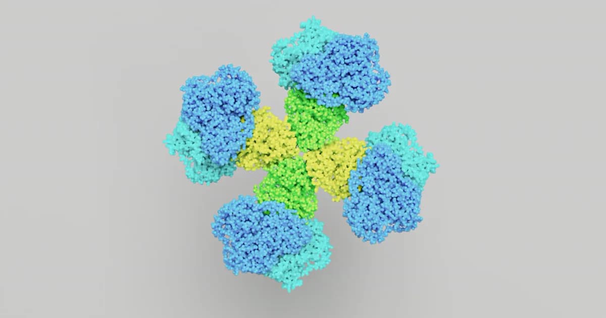
Two Peptides Behind a Familiar Puzzle
Physiologists noticed something odd decades ago. Swallow a glass of sugary juice and your pancreas releases far more insulin than it does when the identical amount of glucose goes straight into a vein. Same sugar. Very different insulin response. The gap comes down to a pair of small gut peptides — GLP-1 and GIP — that together make up the incretin peptide class. The compounds discussed here are offered for research use only, and what follows is a structural explainer, not medical guidance.
For anyone researching this family, the native peptides are the map. A whole generation of engineered research analogues borrows directly from their sequences, their receptors, and — just as importantly — their weaknesses. Understand the two natural incretins and the designed compounds suddenly become legible. This piece walks through what actually earns a peptide the "incretin" label, how GLP-1 and GIP are built, the receptors they dock into, why they fall apart so quickly, and how that fragility drives everything researchers construct next.
What Makes a Peptide an "Incretin"?
Start with the practical question: what does the word actually mean? An incretin is a hormone released from the gut after eating that prompts the pancreas to release insulin — but only when blood glucose is already elevated. That last clause carries the weight. The signal is glucose-dependent, which is why researchers describe these peptides as glucose-dependent insulinotropic agents rather than blunt insulin triggers.
This is the mechanism behind the juice-versus-IV puzzle. Glucose arriving through the digestive tract prompts the gut to secrete incretins that amplify the insulin response; the same glucose given intravenously bypasses the gut and skips that amplification. The gap between the two is what researchers call the incretin effect, and in cell and tissue studies it accounts for a large share of the insulin released after a meal, as reviewed in the classic biology of incretins literature.
In humans the class has exactly two members: glucagon-like peptide-1 (GLP-1) and glucose-dependent insulinotropic polypeptide (GIP). They are grouped together by what they do, not by how they are spelled — the two peptides come from entirely different genes and share only modest sequence overlap. What they do share is a structural habit: each folds into an alpha-helix as it binds its receptor, a shape researchers can read with tools like circular dichroism. That helix is the working end of the molecule.
GLP-1 Structure: A Fragment of Proglucagon
Origin and processing
GLP-1 doesn't have its own dedicated gene. It's carved out of a much larger precursor called proglucagon, and which fragment you get depends on the tissue doing the cutting. In pancreatic alpha cells the enzyme prohormone convertase 2 processes proglucagon into glucagon; in intestinal L-cells and certain neurons, prohormone convertase 1/3 processes the same precursor into GLP-1 and its neighbors, as detailed in the GLP-1 structural overview. Same starting protein, different scissors, different product.
The two active forms
Mature GLP-1 is a compact 30- or 31-residue peptide, and it circulates as two forms that are essentially equal in potency: GLP-1(7-36)amide and GLP-1(7-37). The naming refers to where the active fragment begins and ends within the precursor. The amidated version — capped at its tail by an amide group rather than a free acid — predominates, making up more than 80% of what is secreted. That terminal chemistry is no footnote; whether a peptide ends in an amide or a free acid changes its stability and behavior, a distinction worth understanding through the lens of C-terminal amidation.