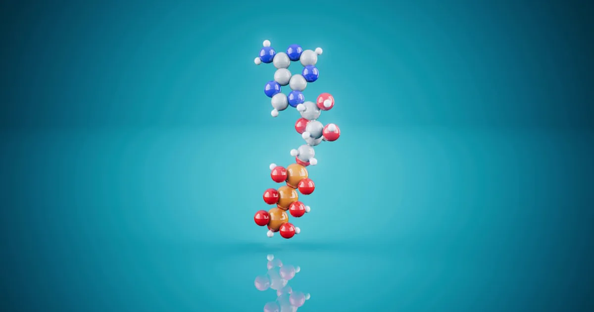
PEGylation Chemistry: How PEG Conjugation Changes a Peptide's Properties
PEGylation bolts an inert polymer onto a peptide and quietly rewrites how the body handles it — slowing clearance, boosting solubility, and shielding it from enzymes. Here's the chemistry behind those changes, and the trade-offs researchers weigh when they choose where PEG attaches, how big it is, and what linker holds it on.
by Research Assistant·
A peptide can be a beautifully designed molecule and still fail on the bench because it vanishes from circulation within minutes. The information here is offered for research use only, to explain a chemistry problem researchers hit constantly: many peptides are potent but short-lived, cleared by the kidney and chewed up by enzymes almost as fast as they appear. PEGylation is one of the most common ways to change that. It doesn't rewrite the peptide's sequence — it attaches a chain of polyethylene glycol (PEG), an inert, water-loving polymer, and lets that chain do the work.
So what actually happens to a peptide's properties when you bolt PEG onto it? This piece walks through the core mechanism behind the longer half-life, the other physical changes PEG brings, and the three levers — where PEG attaches, how big it is, and what linker holds it on — that turn a blunt "add polymer" step into precise molecular engineering.
Why Researchers Reach for PEG in the First Place
The short version: small peptides are powerful but fragile. Their small size means the kidney filters them out quickly, and their exposed backbone makes them easy targets for proteases. Both problems shorten the window in which a peptide can act.
PEG is a convenient fix because of what it isn't: chemically inert, extremely water-soluble, and only weakly recognized by the immune system. Attaching it changes a peptide's behavior in circulation without touching the amino acid sequence that gives the peptide its function. That makes PEGylation one entry in a broader toolkit of stability strategies — researchers might instead consider swapping in D-amino acids to resist enzymatic breakdown, for example. What sets PEG apart is that it changes pharmacokinetic and physical properties — how long the molecule lasts, how well it dissolves, how visible it is to enzymes — rather than altering the peptide's own chemistry.
How PEG Extends Circulation Time
The single biggest thing PEG does is make the molecule "look bigger" to the kidney. Everything else follows from that.
The hydrodynamic radius mechanism
Kidney filtration depends on hydrodynamic size, not just raw mass — as one review puts it, plasma half-life is a function of hydrodynamic size. PEG inflates that size out of proportion to its weight because it drags a large shell of water with it. Each ethylene oxide unit in the chain binds two to three water molecules, wrapping the peptide in a hydrophilic cloud. The effect on apparent size is striking: salmon calcitonin modified with a PEG5000 chain behaved as though it were 259 kDa, far larger than its actual 84 kDa mass. To the kidney's filter, the conjugate simply reads as too big to pass through quickly.
What that buys, in numbers
The payoff is measured in hours and days rather than minutes. Conjugating a drug to a roughly 40 kDa PEG has been used with peptides, nucleic acids, and small molecules and can afford half-lives of up to about seven days in humans. On the protein side, pegfilgrastim shows roughly a ten-fold longer half-life than unmodified recombinant human G-CSF. Same molecule, same active site — a very different residence time in circulation.
Beyond Half-Life: Solubility, Stability, and Immune Shielding
Slower clearance is the headline, but PEG changes a peptide's physical behavior in three other practical ways.
Solubility
That same water-loving shell that fools the kidney also pulls the conjugate into solution. For peptides that tend to clump or fall out of solution, a PEG chain can raise aqueous solubility and make the material easier to handle.
Proteolytic resistance
PEG acts like a physical screen. By draping over the peptide surface, it blocks proteases from reaching the bonds they would otherwise cut. PEGylation of the antimicrobial peptide nisin A, for instance, substantially enhanced its enzymatic resistance. The peptide is the same; it's simply harder for enzymes to get a grip.
Lower immunogenicity
The same shielding lowers immune recognition. Hide the surface features an antibody would latch onto, and you blunt the immune response — with the enzyme asparaginase, PEGylation could eliminate its antigenicity. For a molecule meant to persist in circulation, staying invisible to the immune system is part of staying active.
Where PEG Attaches: The Conjugation Site
Chemists don't just add PEG — they choose which atom it bonds to, and that choice shapes the entire product. A handful of reactive groups on a peptide serve as the anchor points.
N-terminal amines
The amino group at a peptide's N-terminus can be targeted selectively by exploiting a difference in acidity: the N-terminal amine has a pKa around 7.6 to 8.0, while lysine side chains sit near 10.0 to 10.2. Run the reaction at mildly acidic pH and PEG reacts at the terminus while the lysines stay largely untouched.
Lysine amines
Lysines are the easy, abundant target — and that's the problem. Because a protein usually carries many of them, coupling to lysine amines tends to produce a heterogeneous mixture of products with PEG in different places, which is hard to purify and characterize.
Cysteine thiols
Free cysteines are comparatively rare, which makes them a precise handle. Maleimide chemistry reacts cleanly with cysteine's thiol group, so if a peptide has one available cysteine, PEG can be steered there almost quantitatively without disturbing the disulfide bonds that hold the fold together.
Carboxyls and engineered handles
Glutamate and aspartate carboxyl groups offer another route, activated under acidic conditions with a coupling reagent such as EDC. And when a peptide lacks a convenient natural site, chemists engineer one in — the same logic as deliberately modifying a peptide's terminus to install exactly the reactive group they want.
Random vs Site-Specific PEGylation
The takeaway here is simple: attaching PEG in one defined spot beats scattering it everywhere. Random amine PEGylation gives positional isomers, a purification headache, and a real risk that PEG lands on or near the active site and blunts activity.
Site-specific PEGylation avoids that by directing the polymer to a single location. A clear example comes from work on TIMP2, a 22 kDa protein. Non-specific amine coupling was efficient but produced multiple positional isomers that were difficult to separate. Directing PEG instead to a C-terminal histidine tag, using PEG-bis-sulfone chemistry, produced defined mono- and di-PEGylated forms. The mono-PEGylated version carrying a 10 kDa PEG showed a 6.2-fold longer circulating half-life — from 68 minutes out to roughly 422 minutes — and remained detectable four days later, while unmodified TIMP2 disappeared within hours. Best of all, it held onto its function: the PEGylated protein kept its MMP-inhibitory activity and showed a circular dichroism spectrum nearly identical to the unmodified protein. Homogeneity plus retained activity is the whole point of site control.
PEG Size and Shape: Linear vs Branched
Here's the tension at the heart of PEGylation: bigger PEG lasts longer but can smother activity, and the polymer's shape is one way to thread that needle. As a rule, smaller PEG chains give shorter plasma half-lives, so there's constant pressure to go larger — yet larger chains sit like a bulky cloud over the peptide and can block the surface it needs to do its job.
The numbers show how sensitive this balance is. A linear 20 kDa PEG on TNF-alpha left it with 58% of its activity, while a branched 10 kDa PEG retained 93% — and pushing to a branched 40 kDa chain caused a loss of activity altogether. Branched architectures tend to shield the peptide from proteases better than linear ones, and in one comparison a two-armed branched PEG gave the longest half-life of the shapes tested. Shape, in other words, is a design variable in its own right — not just a detail of how the chain is made.
The Linker Matters Too
The small chemical bridge between peptide and PEG is easy to overlook, yet it quietly steers where the conjugate ends up in the body. A study comparing maleimide-thiol and DBCO-azide linkers on the same CAQK peptide found that both extended blood half-life by about 90-fold — but they distributed very differently. The hydrophobic DBCO linker drove higher accumulation in the kidney and liver, apparently lingering in the reticuloendothelial system, while the maleimide version cleared more quickly through the kidney. Same peptide, same PEG, different linker — and a measurably different off-target footprint.
Linkers can also be designed to let go. In reversible PEGylation, a cleavable bridge releases the free peptide after it has been delivered, so the active molecule isn't permanently saddled with its bulky carrier. Using tunable beta-eliminative linkers, a circulating PEG conjugate achieved a 56-fold longer half-life for the 39-amino-acid peptide exenatide in rats, with the release rate tunable across a remarkable range — from hours to more than a year — just by adjusting the linker's chemistry.
The Trade-Off: What PEG Costs You
PEG is a tool, not a free upgrade, and it helps to be clear-eyed about the costs. Large or non-specifically placed PEG chains routinely lower a peptide's intrinsic activity, which is why so much of the chemistry above is about clawing that activity back. There are documented downsides at the whole-organism level too: reviews note immune reactions and renal effects tied to the accumulation of PEG metabolites over time. None of this makes PEG a poor choice — it remains one of the most reliable ways to extend circulation — but it does explain why researchers weigh alternatives such as cyclizing the backbone for stability, and why the "best" PEGylation strategy is always specific to the peptide in hand.
Frequently Asked Questions
What is peptide PEGylation in simple terms?
PEGylation is the covalent attachment of one or more chains of polyethylene glycol (PEG) — an inert, water-loving polymer — to a peptide or protein. In research settings the goal is usually to change how the molecule behaves in circulation: PEG makes the conjugate larger and more hydrated, which slows how quickly the kidneys filter it out and shields it from enzymes and immune recognition.
Why does adding PEG make a peptide last longer in circulation?
A small peptide is cleared quickly because the kidney filters molecules below roughly 60 to 70 kDa. PEG carries a large hydration shell — each ethylene oxide unit holds two to three water molecules — so a modest PEG chain gives the conjugate a much larger hydrodynamic radius than its actual mass suggests. That larger apparent size retards renal filtration, which is the main reason a roughly 40 kDa PEG can extend a half-life to as long as about seven days in humans in reported studies.
What is the difference between random and site-specific PEGylation?
Random PEGylation attaches PEG wherever a reactive group is available — typically the many lysine amines on a protein — producing a mixture of positional isomers that is hard to purify and can block the active site. Site-specific PEGylation directs PEG to one defined location, such as an N-terminus, a single cysteine, or an engineered histidine tag, yielding a homogeneous product that is easier to characterize and more likely to retain biological activity.
Does PEGylation lower a peptide's activity?
It can. A large PEG chain sits like a bulky cloud around the peptide and can partially block the surface that binds its target, so intrinsic activity often drops even as half-life rises. In one reported example, a linear 20 kDa PEG on TNF-alpha retained 58% of activity while a branched 10 kDa version retained 93%. Researchers manage this trade-off by tuning PEG size and shape, choosing the attachment site carefully, or using releasable linkers that free the active peptide after delivery.
The Bottom Line
PEGylation doesn't change what a peptide is — it changes how the body handles it, trading a measure of intrinsic activity for dramatically longer, more soluble, less immunogenic time in circulation. The real craft lives in three levers: the attachment site determines how clean and active the product is, PEG size and shape set the balance between longevity and function, and the linker steers where the conjugate goes and whether it eventually releases its cargo. Pull those levers well and a fragile, fast-cleared peptide becomes a far more durable research tool. For a closer look at how these modifications interact with the peptide underneath, the related structural-chemistry explainers on cysteine bridges and backbone cyclization are a good next stop.
For research use only. Not for human or animal
consumption of any kind. The information in this article is for
educational purposes only and is not intended to diagnose, treat,
cure, or prevent any disease. The statements made have not been
evaluated by the U.S. Food and Drug Administration. These products
are NOT FDA APPROVED. Please consult with a licensed healthcare
professional before making any decisions regarding your health
or research.
Optides LLC is a chemical supplier. Optides LLC is not a
compounding pharmacy or chemical compounding facility as defined
under 503A of the Federal Food, Drug, and Cosmetic Act. Optides LLC
is not an outsourcing facility as defined under 503B of the Federal
Food, Drug, and Cosmetic Act.
Tags
PegylationPeg ConjugationHalf Life ExtensionPeptide ModificationResearch Peptides
Stay In The Loop
Stay Updated on New Research Compounds
Be the first to know about new products, research guides, and exclusive offers.